Facilities
We have multiple laboratories and equipment to assist with our soft matter, radiation and medical physics, photonics and quantum sciences and experimental nuclear physics research.
Take a tour of our physics facilities
Take a virtual tour around some of our leading physics facilities, including the Physics Undergraduate Labs, Advanced Technology Institute and the Observatory.
Experimental nuclear physics facilities

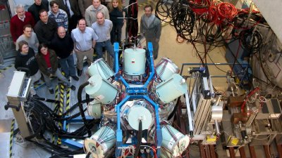
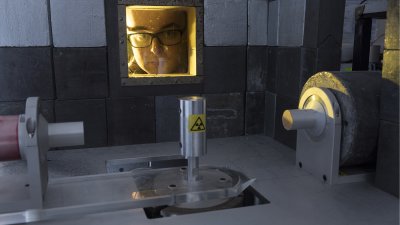
Here at Surrey detection systems are tested and developed and a suite of heavily shielded gamma-ray (hyper pure germanium) detectors for low-level counting of environmental radioactivity is installed.
Facilities we use around the globe
Our Nuclear Physics Experimental Group wins beam time to perform experiments at accelerator laboratories around the world such as gamma-ray experiments using the RISING and EURICA detector arrays at GSI/FAIR (Germany) and RIKEN (Japan) and reactions induced by radioactive beams at TRIUMF (Canada) and CERN/ISOLDE (Geneva).
High performance computing (HPC) clusters
The Astrophysics, Nuclear Theory, Photonics and Quantum Sciences, and Soft Matter groups all use the University's central HPC cluster: Eureka. Eureka is made up of approximately 100 compute nodes. These nodes include some GPU and high-memory nodes. In total, the cluster provides 2200+ CPU cores and 14.5+ TB of RAM. All nodes are based on Intel Xeon CPUs. The cluster runs a mixture of InfiniBand and Omni-Path as high-speed interconnects. The University also has a Condor-based GPU-server farm.
We also have another HPC cluster in the Advanced Technology Institute: Kara, which has 40 compute nodes, including some GPU and high-memory nodes. In total, the cluster provides 1100+ CPU cores and 6.5+ TB of RAM. All nodes are based on Intel Xeon CPUs. This cluster runs InfiniBand as its high-speed interconnect.
Soft matter laboratories

Our Department’s experimental soft matter research by the Soft Matter Group is conducted in recently refurbished laboratories. They have:
- A contact angle analyser (used to study the shape of drops of liquids on a surface, as a means to determine the surface tension)
- 3D printer (used for conventional polymers and also for natural macromolecules that are used in foodstuffs)
- Discovery HR-2 rheometer (capable of variable frequency measurements at varying temperatures)
- A suite of thermal analysis techniques including dynamic scanning calorimetry, dynamic mechanical, thermal mechanical and thermogravimetric analysis.
Microscope and spectrometers
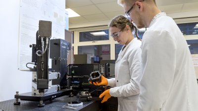
We have both a combined atomic force microscope (AFM) and raman spectrometer and a conventional AFM. Our NTEGRA spectra combined AFM and raman spectrometer is a unique integration of scanning probe microscope and confocal microscopy luminescence and raman scattering spectroscopy.
Ellipsometry
The labs have a variable angle spectroscopic ellipsometer. Ellipsometry provides information about the thickness of surface layers, the complex refractive index, and surface roughness. Our J.A. Woollam ellipsometer offers wavelengths ranging from the ultraviolet (200 nm) to the near infrared (2500 nm). It is equipped with a heating stage and a liquid cell.
Nuclear magnetic resonance/magnetic resonance imaging facilities
We also house a suite of six 1H time domain nuclear magnetic resonance / magnetic resonance imaging facilities for the characterisation of 1H bearing fluids in porous media (e.g. wood and cement). Sample sizes vary from 0.1 to 10 cm; spatial resolution from micrometres to millimetres and pore sizes accessed can be down to nm – although not all at the same time, of course.
Radiation and medical physics facilities

The Radiation and Medical Physics Group run our Department’s ionising radiation detector processing and characterisation laboratories.
Detector preparation laboratories
The detector preparation laboratories allow us to do wet chemistry, cutting, polishing, metallisation, photolithography, annealing, and surface profile measurements.
Characterisation laboratories
Radiation detector characterisation uses nuclear electronics and signal processing, including digital; with standard radio-isotope sources and variable temperatures (100 to 500 K).
Electrical characterisation includes current voltage, capacitance voltage measurements, and photoinduced current transient spectroscopy. The main optical techniques employed are:
- Photoluminescence and photo current measurements
- Optical microscopy, including birefringence measurements during detector operation.
The group’s X-ray imaging research includes micro- CT and phase contrast imaging.
