Head Space Project (Canine Chiari Group)
Start date
September 2021End date
September 2023Project website
ViewBackground
Brachycephaly is associated with many health and welfare problems. One of the most serious is Chiari-like malformation (CM). CM is characterised by a short skull resulting in brain and upper spinal cord overcrowding causing pain and leading to spinal cord cavities called syringomyelia (SM). It is common - one UK referral practice found that 9.7% of all dogs presented with neck pain had CM (CM-P) or SM. SM can result in neurological problems include debilitating “phantom” scratching, scoliosis and weakness.
Currently SM is diagnosed by MRI whereas a diagnosis of CM-P is made by a combination of appropriate history and clinical signs, exclusion of other causes of pain and MRI. In some breeds, for example Cavalier King Charles spaniels (CKCS) and Chihuahua, CM is so common that it is very challenging to find an unaffected dog.
We know that certain head shapes make CM-P and SM more likely – dogs with a higher cephalic index (broad and short head) with a prominent stop and reduced muzzle appear more at risk. This means breeders could select for a less risky head shape if they knew what to select for.
Aims and objectives
In the Head Space Project, we aim to create an objective assessment of head shape using facial recognition technology to better define the face of CM-P and SM. To achieve this, we first built a dedicated 5-camera system which can gather 3D data to generate a 3D model of the complete head. We are now making 3D computer models of dogs with known clinical and MRI CM-SM status. We are comparing 3 groups of CKCS: 1) clinically normal CKCS – so-called Chiari malformation – normal (CM-N) 2) CKCS with clinical signs from syringomyelia – so called syringomyelia – severe / signs (SM-S) 3) CKCS with clinical signs of pain and no syringomyelia – so-called Chiari malformation pain (CM-P)
Progress in so far
Development of the system
Thanks to the skills of Dr Mehran Taghipour Gorjikolaie the five-3D camera system was developed and modified so that it could successfully capture the entire head in less than a second. This was an important development because even when a dog sits on command, they often move their head.
Pilot studies with University of Surrey teaching dogs
The University of Surrey teaching dogs are pets owned by staff and students and several of them participated (in return for treats!) in the pilot study. Following this Mehran rewrote and adapted computer code to improve the system.
Imaging of CKCS at the Companion Cavalier Group AGM
The Canine Chiari Group were delighted to be invited to this event and following a lecture by Clare, all the 21 CKCS at the event in May were successfully imaged.
Imaging of CKCS at Bliss Cavalier Rescue
Sadly, many CKCS entering rescue are affected by CM-P and SM and consequently many have had neurological assessment and MRI and (thanks to Cavalier Matters Charity) in many cases CT. Thanks to the hard work of volunteers at Bliss Cavalier rescue - the Canine Chiari group were able to image 24 dogs that attended this event in October. These dogs also had a respiratory function grading to contribute to the BOAS research under Dr Jane Ladlow at Cambridge university.
Funding amount
£134,563
Funder
Dogs Trust
Team
Principal investigators

Professor Clare Rusbridge
Professor in Veterinary Neurology
Biography
A veterinary neurologist who has spent over 25 years researching Canine Chiari malformation and syringomyelia.

Professor Kevin Wells
Professor of AI in Human and Veterinary Healthcare
Biography
Reader in Medical Imaging and an expert in state-of-the-art computer vision technology and machine learning.
Co-investigator
Additional members

Emma Theobald
Research Fellow at University of Surrey’s School of Veterinary Medicine
Biography
A Research Fellow with vHive at University of Surrey, spending most of her time researching Health Related Quality of Life (HRQoL) in canine and feline osteoarthritis and other canine osteoarthritis research. However, she very kindly donates personal time and expertise to the Canine Chiari Group.

Jake Cumber
Postgraduate Researcher at Centre for Vision, Speech and Signal Processing at University of Surrey
Biography
Jake was originally trained in mathematics, gaining a First Class Integrated Master’s degree in Mathematics from the University of Kent. Now he is a postgraduate researcher at the University of Surrey, working towards a PhD on applications of AI in Canine Chiari and syringomyelia.
Complementary projects
The Canine Chiari group has several simultaneous and complementary projects – one of these is:
Improving screening for CM-P and SM using MRI and CT
Breeders are advised to screen dogs for SM status prior to breeding but there is no robust system for categorising CM and risk of CM-P. Better characterisation of a healthy versus unhealthy skull shape is urgently required so that vigorous and economic screening schemes are possible.
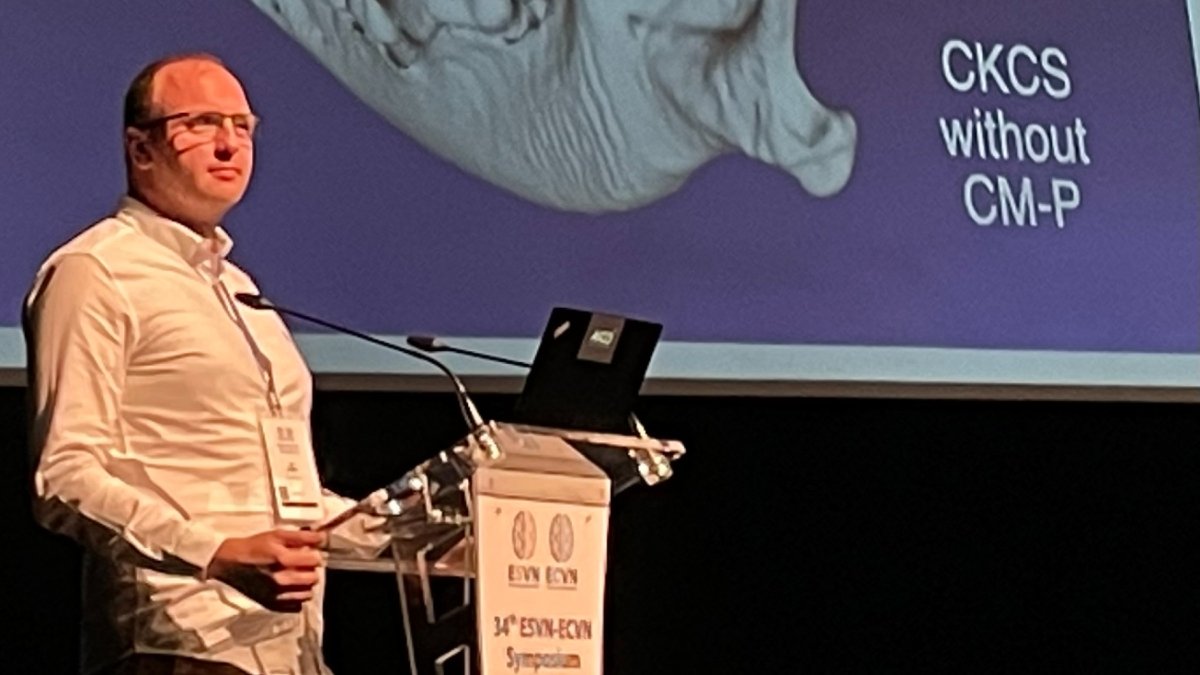
Jake Cumber’s doctoral project uses a machine learning technique to grade MRI scans for SM and CM-P using artificial intelligence. His research won the best neuroimaging presentation at the 2022 European College of Veterinary Neurology Annual Symposium.
The current BVA CM/SM health scheme relies on a single, small field of view, one-dimensional MRI and therefore cannot represent 3D morphological changes of the skull and cervical vertebrae. Compared to MRI, CT is a significantly lower cost screening technique with full 3D data. In the future Jake hopes to develop his novel machine learning based on spherical harmonics to CT and 3D MRI in CKCS and Chihuahua in the anticipation that this could be a useful and more economic screening test for breeders
Project images
Research themes
Find out more about our research at Surrey:


