Lab-grown pig heart tissue could help replace live animals in heart disease research
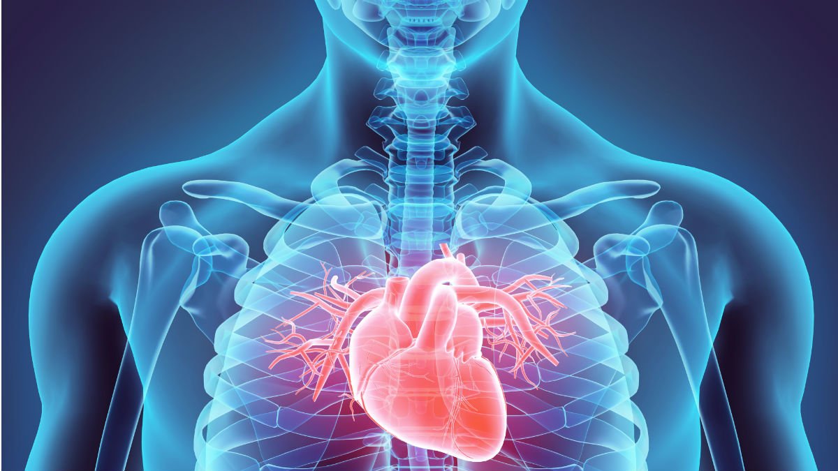
A new way to replicate what happens inside the heart after cardiac arrest could open new avenues for the study of heart regeneration whilst reducing the use of live animals in research, according to a study from the University of Surrey and King’s College London.

Researchers have developed a process that involves obtaining and growing thin slices of pig heart tissue which include both the epicardium --- the most external layer of the heart that contains cells that can promote its recovery --- and underlying heart muscle.
The team treated the epicardial slices with stimulating compounds, showing that cells become activated in a way that replicates what happens in the heart after a heart attack. The new process was able to reproduce observations typically obtained in live animal models.
Dr Paola Campagnolo, lead author of the study and Senior Lecturer in Molecular Cardiovascular Sciences at the University of Surrey, said:
“This research typifies the One Health, One Medicine ethos at the University of Surrey, as our model could help us understand how to stimulate the repair process after heart attacks without the need to use live animals in the research. We are hopeful our model could lead to better health outcomes for humans and reduce the reliance on animal experiments in cardiovascular science.”
According to the British Heart Foundation, there are around 7.6 million people living with heart or circulatory disease in the UK. This disease causes a quarter of all deaths in the UK.
The ability of the heart to recover after an injury is severely limited by the low number of regenerating cells within its tissue. The current research models and strategies aimed at improving the heart’s repair process are mainly based on surgical procedures performed on laboratory animals.
Dr Davide Maselli, Postdoctoral Research Associate and first author of the paper, said:
“This work provides an innovative tool to study the healing from a heart attack. We believe that our model could be useful to dissect the role of different cells in the reparative process. In our consideration, it is extremely important that every step forward in this field delivers a clinical perspective for the patients while reducing the burden on research animals.”
The research, published in the journal npjRegenerative Medicine, proposes a system to study the regeneration of the heart in a laboratory dish and could therefore lead to a reduction in the number of small animals used in cardiovascular research. It is estimated that the implementation of the model could reduce the use of animals in this type of research by 20%. A typical preliminary study uses 20 to 40 rodents, and a lab can perform several of these studies each year. A replacement of 20% could equate to as much as 1,000 fewer animals required for epicardial studies per year in the UK.
Notes to Editors:
This work was funded by the NC3Rs (National Centre for the Replacement, Refinement & Reduction of Animals in Research).
Epicardial slices: an innovative 3D organotypic model to study epicardial cell physiology and activation can be found here.
Dr Paola Campagnolo is available for interview upon request.
University of Surrey Media Team contact details: mediarelations@surrey.ac.uk
Media Contacts
External Communications and PR team
Phone: +44 (0)1483 684380 / 688914 / 684378
Email: mediarelations@surrey.ac.uk
Out of hours: +44 (0)7773 479911
